Juan Pinto, MD, FAAD


Board-Certified Dermatologist
Fellowship-Trained Mohs Surgeon
Fellow of the American Academy of Dermatology


Dr. Juan M. Pinto is a board-certified dermatologist and fellowship-trained Mohs surgeon specializing in skin cancer treatment and advanced facial reconstruction. Born and raised in Venezuela, he is a fifth-generation physician and completed formal training in plastic and reconstructive surgery before moving to the United States. He earned his medical degree from Universidad Central de Venezuela and later completed his dermatology residency and Micrographic Surgery and Dermatologic Oncology (Mohs) fellowship at University of Texas Medical Branch.

Dr. Pinto is known for his meticulous surgical approach and expertise in Mohs surgery and complex facial reconstruction, with a focus on achieving optimal functional and aesthetic outcomes. He is board-certified by the American Board of Dermatology and is an active member of several professional organizations, including the American Academy of Dermatology and the American College of Mohs Surgery.

Outside of practice, Dr. Pinto enjoys spending time with his wife, Ayezel, and their daughter, Paula, as well as traveling, cooking, and exploring diverse cuisines. He provides specialized Mohs surgery and reconstructive care in the Houston area.

Additional Languages
  • LanguageIcon Spanish
Specialties and Affiliations
  • American Academy of Dermatology
  • American Society for Dermatologic Surgery

Clinic Locations

U.S. Dermatology Partners Medical District

1213 Hermann Dr
#650
Houston, Texas 77004

(713) 528-8882

Services Offered By Juan M. Pinto, MD

What Is Actinic Keratosis?

Actinic Keratosis, also known as solar keratosis, is a scaly or crusty lesion on the skin that develops slowly and indicates the presence of sun damage. It is most commonly found on parts of the body frequently exposed to the sun including the bald scalp, face, ears, lips, backs of the hands or forearms, neck, and shoulders.

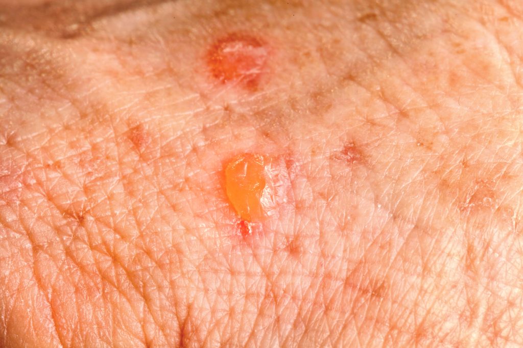
Actinic Keratosis two days after a freezing removal treatment

Actinic keratosis improves just two days after a freezing removal treatment.

Actinic keratoses are considered precancerous and can develop into a type of skin cancer called squamous cell carcinoma. In fact, some 40 to 60 percent of squamous cell skin cancers begin as untreated actinic keratoses.

Because of this, your doctor should be diligent in diagnosing, treating and monitoring actinic keratosis.

What Is Basal Cell Carcinoma?

Basal Cell Carcinoma, also known as basalioma or basal cell cancer, is the most common type of skin cancer and carries the least amount of risk, though it still requires attention. If caught and treated early, basal cell carcinomas are not likely to be life-threatening, but they do have the potential to cause disfigurement of the skin tissue.

Almost one million new cases of basal cell carcinoma are diagnosed each year in the U.S., and up to 30% of Caucasians may develop basal cell carcinomas in their lifetime.

Basal cell carcinoma treatment

Basal cell carcinoma can be treated by removing the affected area.

Skin cancer is considered low risk when the affected cells remain clustered in a single group. Both basal cell carcinoma and squamous cell carcinoma are rarely life-threatening. Though it is unlikely to spread to other parts of your body, if left untreated, basal cell carcinoma can move into nearby bone or other tissue.

Basal cell carcinoma typically begins as a small, shiny bump on the face, although it can occur on any part of the body.

What Is Cryotherapy?

Cryotherapy, or “cryosurgery,” is a simple, non-invasive procedure in which liquid nitrogen is used to freeze and destroy growths on the surface of the skin. This is an effective treatment for precancerous skin lesions (actinic keratoses), as well as other skin conditions such as warts, skin tags and moles.

Applying liquid nitrogen to skin lesions allows dermatologists to target the damaged skin cells and destroy them at the cellular level. After freezing, the affected area may blister and scab over, and should heal within three to six weeks.

Our dermatology team uses cryosurgery to treat a wide range of conditions. It offers a number of advantages: Cryotherapy is a simple, affordable outpatient procedure, the discomfort level is minimal, and there is a low risk of infection.

In cryotherapy treatment, liquid nitrogen is applied to the skin to freeze and destroy the affected tissue.

In cryotherapy treatment, liquid nitrogen is applied to the skin to freeze and destroy the affected tissue.

What Is Melanoma?

Melanoma, the deadliest of skin cancers, only accounts for about 4 percent of all skin cancer cases, but causes about 79 percent of skin cancer deaths.

Melanoma is a cancer of the skin that begins in the melanocytes, which are the cells that produce the pigment melanin. It is the leading cause of cancer death in women 25 to 30 years old and the second leading cause of cancer death in women 30 to 35 years old.

In some cases, melanoma occurs in melanocytes throughout the body, even if those parts have never been exposed to the sun.


What is Merkel Cell Carcinoma?

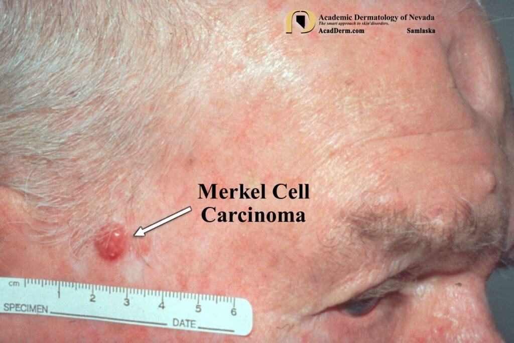
Skin cancer is a common concern in the U.S.  Estimates reported by the American Academy of Dermatology suggest one in five people in the U.S. will develop some form of skin cancer. Merkel cell carcinoma is among the rarest forms of skin cancer, wherein estimates by the Skin Cancer Foundation suggest that only one in 130,000 people in the U.S. will be diagnosed with Merkel cell carcinoma. Like melanoma, Merkel cell carcinoma is an aggressive form of skin cancer with a high mortality rate.  As with most potentially aggressive malignancies, early detection is the best way to decrease the risk of death associated with Merkel cell carcinoma. You can learn more about diagnosis and treatment options for Merkel cell carcinoma on this page.

Merkel cell carcinoma is a rare form of skin cancer that arises from Merkel cells, cells that reside deeper in the skin and function to send ‘touch’ signals from outside the skin to the inside of the body.  The diagnosis of Merkel cell carcinoma is ultimately made by an assessment under the microscope after a skin biopsy has been performed.  Skin biopsies are interpreted by dermatopathologists, doctors who specialize in evaluating skin under the microscope.  Dermatopathologists are well equipped to make this diagnosis accurately when skin tissue is submitted in a biopsy specimen.  Unfortunately, when Merkel cells become cancerous, they also become aggressive.  Merkel cell carcinoma can metastasize (spread) quickly, making it one of the most aggressive types of cancer.  While treatment for Merkel cell carcinoma may be successful, even with treatment Merkel cell carcinoma may evade what appears to be disease remission, and recur.


What Is Mohs Surgery?

Mohs surgery offers the highest cure rates for all non-melanoma skin cancers. For certain cases of the most common types of skin cancer — squamous cell carcinoma and basal cell carcinoma — the cure rate can be as high as 99 percent.

Mohs surgery is a highly specialized surgical technique used to treat non-melanoma skin cancers in which the surgeon removes all of the visible cancer, plus a small margin of the surrounding healthy tissue and examines it to ensure that all cancer cells have been removed at the time of surgery.

Before and After Mohs Surgery Technique

The Mohs surgery technique treats skin cancers by removing all of the visible cancer. Image Source: newhealthadvisor.com

During Mohs micrographic surgery — named after Dr. Frederic Mohs, who first performed it in the 1930s — cancer is removed from the skin layer by layer until all cancerous cells have been removed. This type of surgery is most commonly used for cancers that have a high risk of re-occurrence. This technique allows for complete removal of the skin cancer while minimizing the removal of surrounding healthy skin.

What Is Sebaceous Carcinoma?

A rare and aggressive form of skin cancer, sebaceous carcinoma is sometimes referred to as sebaceous gland carcinoma, sebaceous gland adenocarcinoma or meibomian gland carcinoma.

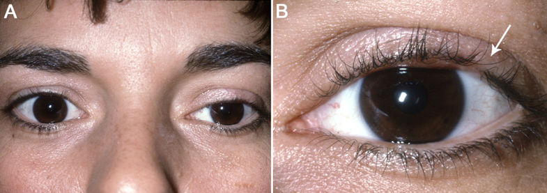
Sebaceous carcinoma can develop in any sebaceous glands, which lubricate the skin, but it most often begins on or around the eyelids. If it is found and treated early, treatment is often successful. However, if sebaceous carcinoma spreads, it can be deadly.

Because sebaceous carcinoma can appear to be a benign growth such as a stye, diagnosis is often delayed, which increases the risk of death. If you notice a growth on your eyelid, it’s important to make an appointment with your dermatologist. The sooner sebaceous carcinoma is diagnosed and treated, the better the outcome.

32-year-old cosmetologist presented with 3-month history of treatment for presumed chalazion. A) The left upper eyelid showed 2 mm of ptosis with swelling. B) Focal loss of eye lashes was appreciated (arrow).” (source: “Early Onset of Sebaceous Carcinoma” by Dr. Debashis Ghosh, creative commons license)

What Is Skin Cancer?

Skin cancer is the most common form of cancer in the U.S. with more than 3.5 million cases diagnosed each year.

Skin cancer is the result of uncontrolled growth of abnormal skin cells that takes place when skin cells suffer DNA damage and then mutate, causing them to multiply rapidly and form malignant (cancerous) tumors. Most skin cancers develop on the visible outer layer of the skin (the epidermis), particularly on sun-exposed areas such as the face, head, hands, arms and legs. They are usually easy to detect with a skin examination, which increases the chances of early diagnosis.

There are different types of skin cancer, each named for the type of skin cell from which they originate. The most common type of skin cancer is basal cell carcinoma. Almost one million new cases of basal cell carcinoma are diagnosed each year in the U.S. Most skin cancers fall into one of three categories:

There are often warning signs that cancer is developing. The most common are pre-cancerous lesions called actinic keratoses that often develop on sun-exposed areas. These tumors replace normal surrounding tissue and generally do not spread to other areas.

Skin cancer is considered low risk when the affected cells remain clustered in a single group. Both basal cell carcinoma and squamous cell carcinoma are rarely life-threatening.

Skin cancer is considered a high risk when cells have invaded surrounding tissues. The third most common skin cancer, malignant melanoma, can be life-threatening if not diagnosed and treated early.

If skin cancer is detected before it has spread to surrounding tissues, the chances of a complete recovery and cure are excellent. High-risk forms of cancer like melanoma require more aggressive treatments.

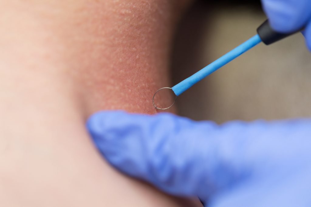
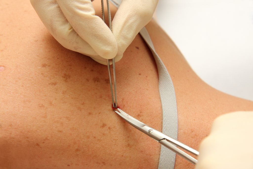
Doctor removing mole from a patient's shoulder.

Suspicious moles should be watched closely, as they might indicate a cancerous growth.

What Is Squamous Cell Carcinoma?

Squamous Cell Carcinoma is a common form of skin cancer that develops in the squamous cells that make up the outer layer of the skin. Although it is usually not life-threatening, it can be aggressive in some cases.

If left untreated, squamous cell carcinoma can grow large or spread to other parts of your body, causing serious complications.

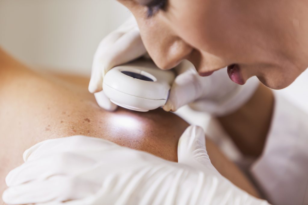
Dermatologist examining male patient's skin for signs of Squamous Cell Carcinoma.

Your dermatologist will be able to examine your skin for signs of squamous cell carcinoma.

What Is a Lipoma?

Lipomas: Understanding & Addressing Benign Fatty Growths

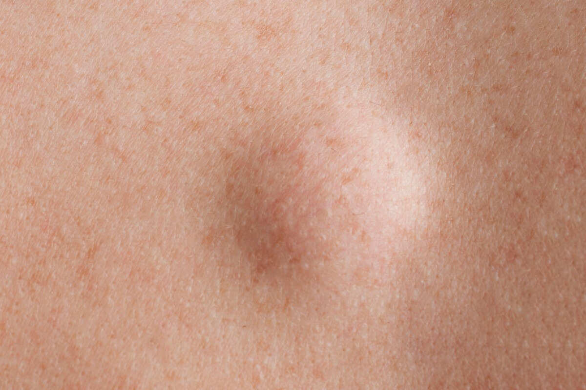
A lipoma is a benign fatty growth, most often located in the deep aspect of the skin (subcutaneous tissue).  These growths arise from the normal fat cells in our subcutaneous tissue and are benign (non-cancerous).  However, there is a very rare form of cancer called liposarcoma, which is a malignant growth from these fat cells.  Lipomas are relatively common, impacting about 1% of people.  However, researchers suspect that lipoma incidence is significantly underreported.

A lipoma is a common, non-cancerous, soft tissue growth just below the skin made up of fat cells. Learn more about lipomas here.

What Our Patients Say

Juan Pinto, MD, FAAD
5 Stars  The staff starting from the front desk, the nurse and Dr. Pinto were wonderful. I was greeted and helped with all my forgetfulness with patients. The nurse was polite, thorough and friendly. Upon meeting Dr. Pinto my positive feelings were even more reinforced. He listened very attentively and wanted me to be completely comfortable with discussing my situation. He then explained thoroughly what should and could be done. I made my decision with confidence because of my positive impressions about him. I could not ask for a more positive experience. Thank you, Terry McFarland –
Source : Google – Jul 10, 2026
Juan Pinto, MD, FAAD
5 Stars  Doing much better. Dr Pinto called to check up on me today. –
Source : Google – Jul 10, 2026
Juan Pinto, MD, FAAD
5 Stars  Dr. Pinto was personable and very thorough and kind. He took time to explain the procedure with me before he did anything. I had a great outcome, would recommend him! –
Source : Google – Jul 09, 2026

Ready to Get Started?

×

How do you prefer to schedule your appointment?

CALL ME LIVE CHAT